Creative Scope + Contributions
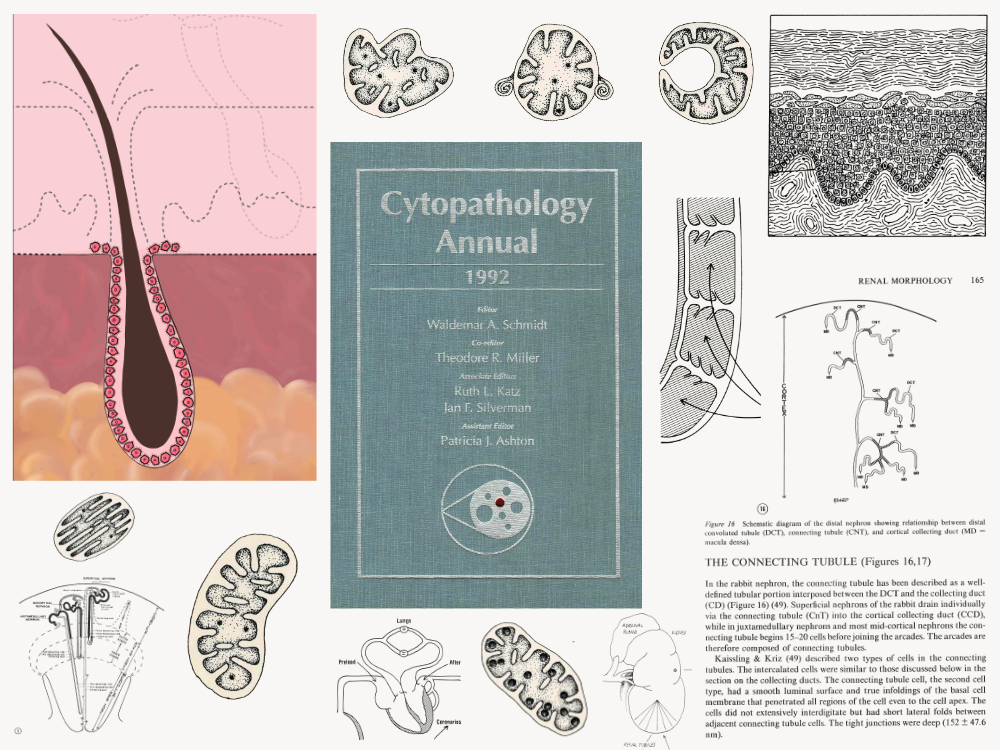
Illustrations for medical publications, textbooks, academic syllabi, and poster sessions covering renal morphology, surgical pathology, and general histology and anatomy for two university medical programs. Work included journal figures, textbook illustrations, a book cover, and course syllabi diagrams. Also produced an animated diagram of keratinocyte structure. The core skill across all of it: identifying which anatomical elements to show, and which to omit, so a complex biological structure reads clearly to a non-specialist viewer.
Tools & Production
Pen and ink renderings for print publications, digital animation for the keratinocyte piece.
